visit: www.infertilitytutorials.com
Procedures

Inseminations (IUIs)

Ultrasound

Ovarian Hyperstimulation

Surgery
  • Surgical Incisions
  • Preoperative Diagnosis
  • Prevention of
    Scar Tissue

  • The OR Team
  • Normal Events
  • Complications

In Vitro Fertilization

Patients review their care
with Dr Eric Daiter

Click here for more video reviews

How Can I help You?

Dr Eric Daiter is a highly regarded infertility doctor with 20 years of experience. Dr. Daiter has personally witnessed which treatments are effective in different situations. If you are having trouble getting pregnant, Dr Eric Daiter is happy to help you (in the office or on the telephone). It is easy, just call us at 908 226 0250 to set up an appointment (leave a message with your name and number if we are unable to get to the phone and someone will call you back).

Availability

"I always try to be available for my patients since I do understand the pain and frustration associated with fertility problems or endometriosis."

Cost

"I understand that the economy is very tough and insurance companies do not cover a lot of the services that might help you. I always try to minimize your out of pocket cost while encouraging the most successful and effective treatments available."

Name:


Phone:


Email (Will be kept private):


How can we help?:


Verify code above:

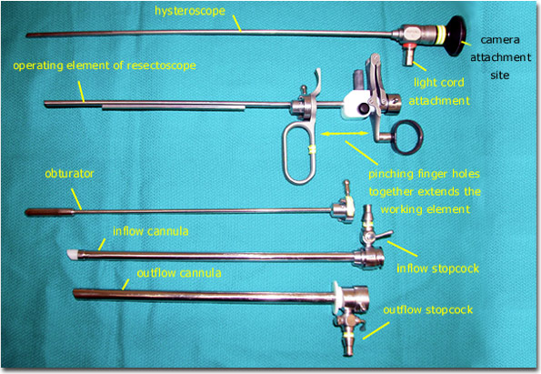
This resectoscope includes 4 main pieces that must be assembled to become functional.

The hysteroscope is passed through the operating element of the resectoscope, locked into place, and a working element (loop electrode, roller ball, etc) is then secured in place within the operating element.

The hysteroscope in this photo appears slightly curved but in fact it is perfectly straight. The apparent curve is a photographic artifact.

A camera can be placed onto the black (viewing) end of the hysteroscope. A light cord is also attached to the hysteroscope.

The working element that is secured to the operating element extends outward when the finger holes of the operating element are pinched together.

The inflow cannula is placed through the outflow cannula and locked into place. The obturator is placed into the inflow cannula to prevent potential blockage by cervical or endometrial tissue during insertion.

The inflow cannula is connected to the distention media at the stopcock via tubing. The outflow cannula is generally connected to a suction device at the stopcock via tubing.


Bookmark This Site  |   Read More Tutorials

The New Jersey Center for Fertility and Reproductive Medicine