|

A number of (types of) ultrasound transducers are currently available. Tranvaginal ultrasound probes contain either mechanical, phased array, or curvilinear transducers that can produce sector images (the displayed image is in the shape of a piece of pie rather than in the shape of a rectangle).
Differences in ultrasound probes are important (to the operator) since they determine display characteristics such as the (1) depth of penetration (as probe frequency increases the depth of visibility or “tissue penetration” decreases and the picture resolution increases... such that as the frequency of the probe increases you see things close to the transducer better but you may not be able to see things farther away); (2) focal zone (some probes will have a fixed focal zone so that only objects that are say 5 cm from the transducer are in focus while other probes will be able to maintain multiple focal zones simultaneously); (3) field of view (this essentially determines “how big a piece of pie” is delivered by the transducer, with an entire circle containing 360 degrees so that a probe with a field of view of 120 degrees is 1/3 of a circle); and (4) resolution (generally structures farther from the transducer are seen with less resolution).
Transvaginal ultrasound probe tips contain the ultrasound transducer (working element) and normally can be placed adjacent to (or at least very close to) the uterus and the ovaries. Therefore, a transvaginal ultrasound probe is ideal in order to obtain high frequency high resolution images of the reproductive organs. The transabdominal ultrasound (jelly on the belly ultrasound) probes are considerably farther from the reproductive organs, the transducer signals need to traverse the (thick) abdominal wall and intervening bowel, so the images (of reproductive organs in the pelvis) are of considerably poorer quality when compared to transvaginal images.
Prior to a transvaginal ultrasound examination, the patient should have emptied her bladder. This is important primarily since a full bladder may occupy most of the pelvis and thereby displaces the ovaries out of the pelvis (farther from the ultrasound transducer).
The transvaginal ultrasound probes are smaller than most vaginal speculums, and therefore, should not be uncomfortable when used during transvaginal ultrasonography. The end of the transvaginal ultrasound probe is covered with an ultrasound coupling gel (to allow better visualization) and then a condom (or protective rubber sheath) is placed over the end of the probe prior to insertion into the vaginal canal. Many ultrasonographers apply a small amount of coupling gel to the outside of the rubber sheath to further enhance the resulting image.
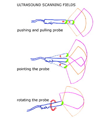
In order to obtain the desired information from the ultrasound exam, the ultrasound probe is pointed in the desired direction, pushed into the vaginal vault to the desired depth for maximal visualization, and rotated to alter the plane of transection.
|

|